


MARK FREI
Formerly of Sigma Aldridge Co.
“When I worked with EBS (while with Sigma-Aldrich), Mike Nesta had a knack for making the difficult look easy. He is extremely bright and his laid back leadership style inspires confidence and gets things done.”
![]()
GREGORY FRITZ
Manager, Microanalysis and Research Software —Thermo Fisher Scientific
“I wanted to say thank you very much for the great support you have provided in regards to the filaments for our LEO 1430.”![]()
MARLON CONTRERAS
Cambridge Vacuum Engineering
“We have worked with other filament manufacturers in the past, but Energy Beam Sciences people understand our business better. Large orders, small orders, they have been very flexible. They are very attentive to our needs!”![]()
MARK ACIERNO
Business Unit Manager, Eurofins Environment Testing Northeast, LLC “Over the last year, we have worked with Energy Beam Sciences to incorporate their DM3250 Microwave Drying Unit into our large environmental testing laboratory. So far, we have been processing three to four hundred samples of soil a day through their technology, sans any mechanical issues. The system is a lot faster than conventional ovens, uses less energy, takes up less of a footprint, and saves manpower. Our resources and operations are more optimized now due to their efficient equipment.”
![]()
![]()
Our Microwave Muffle Furnace is featured on “Metoree”, a product and manufacturer information comparison site for researchers and engineers. Here is the page about Energy Beam Sciences, Inc on Metoree.
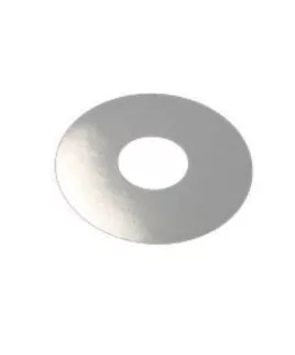
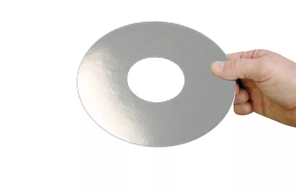
PolarHeat™ 7″ Donut
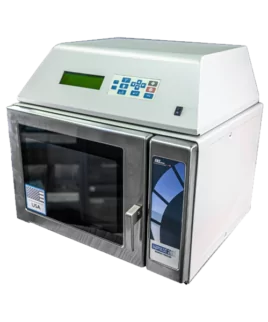
$31.18LabPulse Microwave Tissue Processor
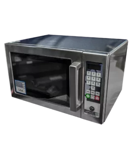
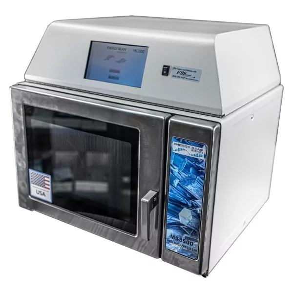
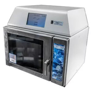
$25,999.00Staining Laboratory Microwave
$5,525.00